Accelerate Biological Research
info@kendallscientific.com
+1-888.733.6849
+1-617.299.7367 (Int’l)
+1-617.299.7367 (Int’l)
+1-888.733.6849
+1-617.299.7367 (Int’l)
+1-617.299.7367 (Int’l)
For quotations, please use our online quotation form, and you may also contact us by
service@kendallscientific.com
+1-888.733.6849 (Toll-free)
+1-617.299.7367 (Int’l))
+1-888.733.6849
Our customer service representatives are available 24 hours, Monday through Friday to assist you.| Reactivity | Human Mouse Rat |
| Tested applications | WB IHC IF |
| Recommended Dilution | WB 1:500 - 1:2000 IHC 1:50 - 1:200 IF 1:50 - 1:200 |
| Calculated MW | 70kDa |
| Observed MW | Refer to figures |
| Immunogen | Recombinant protein of human XRCC6 |
| Storage Buffer | Store at -20℃. Avoid freeze / thaw cycles. Buffer: PBS with 0.02% sodium azide, 50% glycerol, pH7.3. |
| Concentration | ej |
| Synonym | ML8; KU70; TLAA; CTC75; CTCBF; G22P1; |
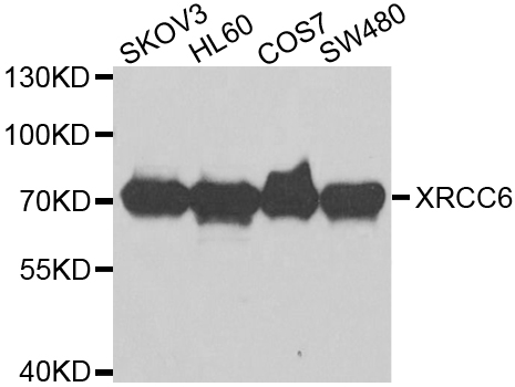
Western blot analysis of extracts of various cell lines, using XRCC6 antibody.
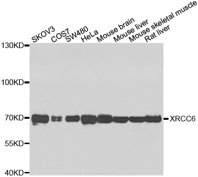
Western blot analysis of extracts of various cell lines, using XRCC6 antibody.

Immunohistochemistry of paraffin-embedded Human adenomyosis using XRCC6 antibody at dilution of 1:100 (x400 lens).

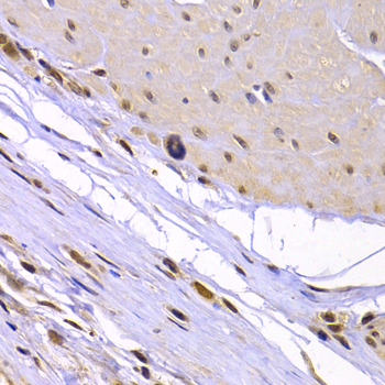
Immunohistochemistry of paraffin-embedded Human brain astrocytoma using XRCC6 antibody at dilution of 1:100 (x400 lens).

Immunohistochemistry of paraffin-embedded Human gastric using XRCC6 antibody at dilution of 1:100 (x400 lens).

Immunohistochemistry of paraffin-embedded Human gastric cancer using XRCC6 antibody at dilution of 1:100 (x400 lens).

Immunohistochemistry of paraffin-embedded Human leiomyoma of uterus using XRCC6 antibody at dilution of 1:100 (x400 lens).

Immunohistochemistry of paraffin-embedded Human mammary gland using XRCC6 antibody at dilution of 1:100 (x400 lens).
Immunohistochemistry of paraffin-embedded human stomach cancer using XRCC6 antibody at dilution of 1:100 (x400 lens).
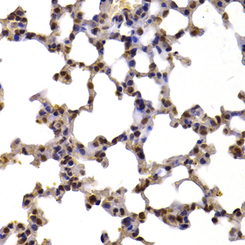
Immunohistochemistry of paraffin-embedded mouse lung using XRCC6 antibody at dilution of 1:100 (x400 lens).
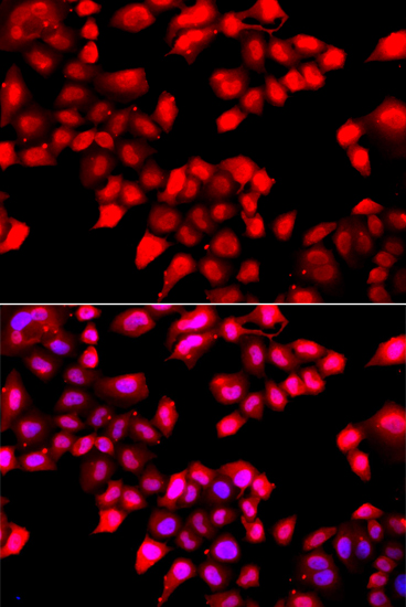
Immunofluorescence analysis of A549 cell using XRCC6 antibody. Blue: DAPI for nuclear staining.
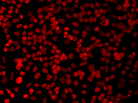
Immunofluorescence analysis of A549 cell using XRCC6 antibody.
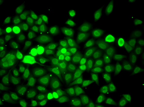
Immunofluorescence analysis of A549 cell using XRCC6 antibody.
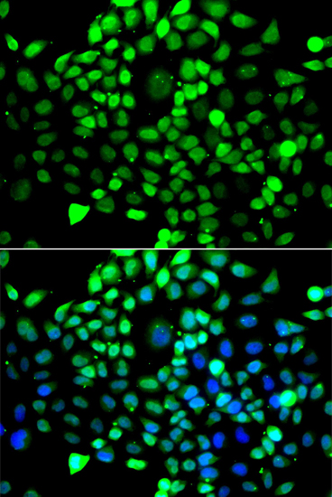
Immunofluorescence analysis of A549 cell using XRCC6 antibody. Blue: DAPI for nuclear staining.
The p70/p80 autoantigen is a nuclear complex consisting of two subunits with molecular masses of approximately 70 and 80 kDa. The complex functions as a single-stranded DNA-dependent ATP-dependent helicase. The complex may be involved in the repair of nonhomologous DNA ends such as that required for double-strand break repair, transposition, and V(D)J recombination. High levels of autoantibodies to p70 and p80 have been found in some patients with systemic lupus erythematosus.
N/A


Copyrights © 2014 Kendall Scientific, 250 Parkway Drive, Suite 150 Lincolnshire, IL 60069


