Accelerate Biological Research
info@kendallscientific.com
+1-888.733.6849
+1-617.299.7367 (Int’l)
+1-617.299.7367 (Int’l)
+1-888.733.6849
+1-617.299.7367 (Int’l)
+1-617.299.7367 (Int’l)
For quotations, please use our online quotation form, and you may also contact us by
service@kendallscientific.com
+1-888.733.6849 (Toll-free)
+1-617.299.7367 (Int’l))
+1-888.733.6849
Our customer service representatives are available 24 hours, Monday through Friday to assist you.| Reactivity | Human |
| Tested applications | WB IF |
| Recommended Dilution | WB 1:500 - 1:2000 IF 1:20 - 1:50 |
| Observed MW | Refer to Figures |
| Immunogen | A phospho specific peptide corresponding to residues surrounding S15 of human p53 |
| Storage Buffer | Store at -20℃. Avoid freeze / thaw cycles. Buffer: PBS with 0.02% sodium azide, 50% glycerol, pH7.3. |
| Concentration | beik |
| Synonym | TP53; P53; LFS1; TRP53; FLJ92943; |
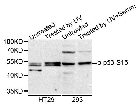
Western blot analysis of extracts of various cell lines, using Phospho-TP53-S15 antibody.
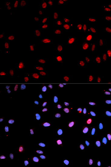
Immunofluorescence analysis of U2OS cell using Phospho-p53-S15 antibody. Blue: DAPI for nuclear staining.
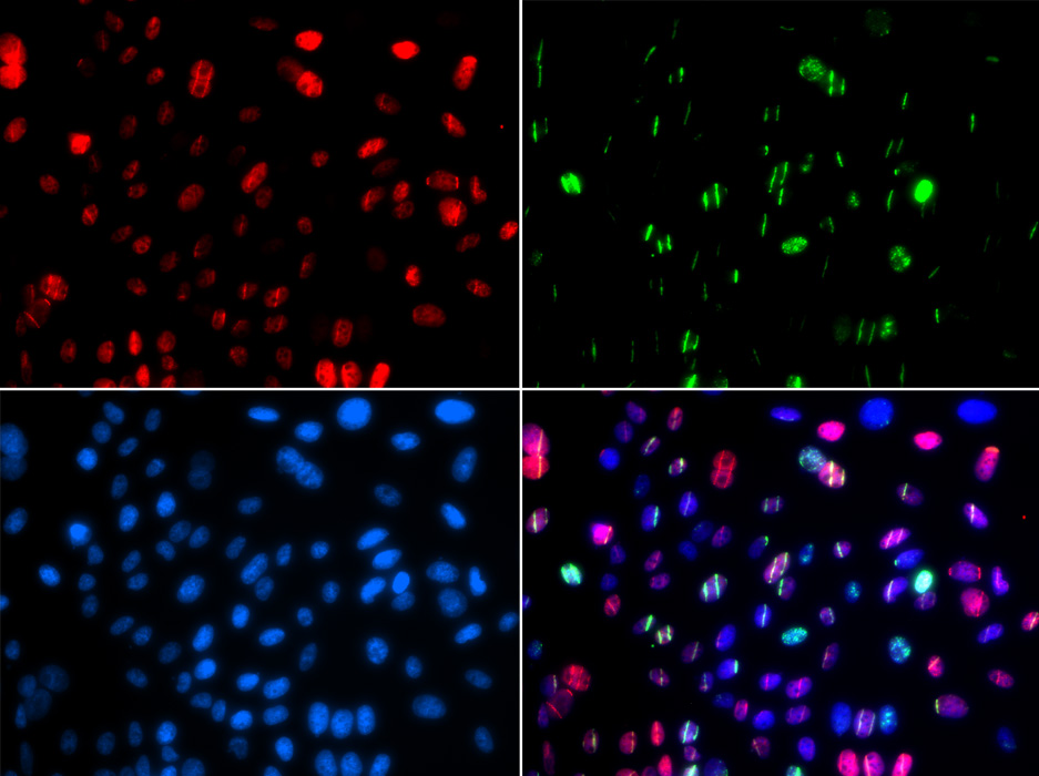
Immunofluorescence analysis of GFP-RNF168 transgenic U2OS cell using Phospho-TP53-S15 antibody. Green:GFP-RNF168 fusion protein expression for DNA damage marker.Blue: DAPI for nuclear staining. RNF168(GFP) can be used to mark cells damaged by UV-A laser for they always gather around DNA damage region.
The p53 tumor suppressor protein plays a major role in cellular response to DNA damage and other genomic aberrations. Activation of p53 can lead to either cell cycle arrest and DNA repair or apoptosis (1). p53 is phosphorylated at multiple sites in vivo and by several different protein kinases in vitro (2,3). DNA damage induces phosphorylation of p53 at Ser15 and Ser20 and leads to a reduced interaction between p53 and its negative regulator, the oncoprotein MDM2 (4). MDM2 inhibits p53 accumulation by targeting it for ubiquitination and proteasomal degradation (5,6). p53 can be phosphorylated by ATM, ATR, and DNA-PK at Ser15 and Ser37. Phosphorylation impairs the ability of MDM2 to bind p53, promoting both the accumulation and activation of p53 in response to DNA damage (4,7). Chk2 and Chk1 can phosphorylate p53 at Ser20, enhancing its tetramerization, stability, and activity (8,9). p53 is phosphorylated at Ser392 in vivo (10,11) and by CAK in vitro (11). Phosphorylation of p53 at Ser392 is increased in human tumors (12) and has been reported to influence the growth suppressor function, DNA binding, and transcriptional activation of p53 (10,13,14). p53 is phosphorylated at Ser6 and Ser9 by CK1δ and CK1ε both in vitro and in vivo (13,15). Phosphorylation of p53 at Ser46 regulates the ability of p53 to induce apoptosis (16). Acetylation of p53 is mediated by p300 and CBP acetyltransferases. Inhibition of deacetylation suppressing MDM2 from recruiting HDAC1 complex by p19 (ARF) stabilizes p53. Acetylation appears to play a positive role in the accumulation of p53 protein in stress response (17).
N/A


Copyrights © 2014 Kendall Scientific, 250 Parkway Drive, Suite 150 Lincolnshire, IL 60069


